
What Is Chicken Bone X Ray and How Does It Work?
chicken bone x-ray technology helps reveal bones hidden inside meat, processed poultry, and prepared foods. It uses controlled X-ray energy rather than cutting the product open. A source sends radiation through the chicken, while a detector records how much energy passes through. Dense bone absorbs more radiation than soft meat. The result is a grayscale image with visible bone edges.
Wilhelm Conrad Röntgen, the pioneer of X-ray science, observed that “the transparency of different substances for the rays is different.” His statement explains the basic contrast behind modern chicken bone x-ray inspection. Today, trained food-safety professionals use calibrated systems, image analysis, and documented procedures to identify fragments. Industrial equipment may inspect products on moving conveyor belts. A small bone can appear as a bright, narrow shape against darker muscle tissue.
The process sounds simple. It is not always simple.
Detection depends on bone size, product thickness, image quality, and the angle of the fragment. Fat, packaging, moisture, and overlapping pieces can also affect contrast. A reliable inspection program therefore combines equipment testing, operator training, maintenance, and careful verification. X-ray inspection does not replace good processing practices. It adds another protective layer.
This article explains what chicken bone x-ray means, how the imaging process works, and why different systems produce different results. It also examines practical limits. Some images look convincing but still need expert review. That uncertainty matters. Clear procedures, validated settings, and responsible interpretation help manufacturers improve safety without promising perfect detection.
What Is a Chicken Bone X-Ray?
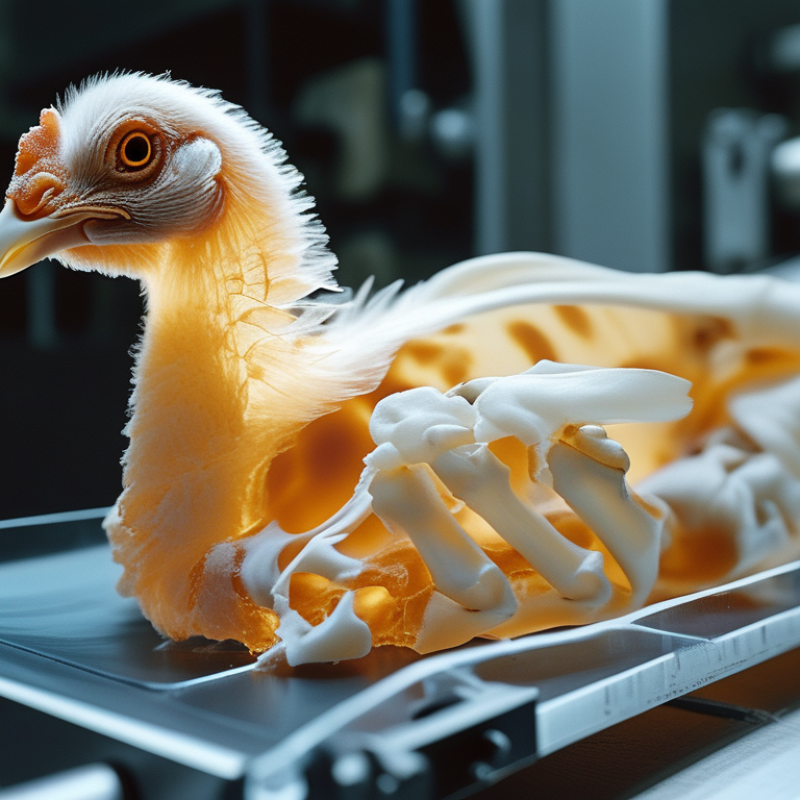
A chicken bone X-ray is a radiographic image used to examine a chicken’s skeleton. It can reveal fractures, abnormal growth, joint changes, or hidden bone damage. During the scan, the bird is carefully positioned while a controlled beam of X-rays passes through its body. Dense bone absorbs more radiation and appears pale on the image. Soft tissues look darker.
The procedure is usually quick, but positioning a live chicken can be difficult. A veterinarian may use gentle restraint or appropriate sedation to reduce movement and stress. Even a small motion can blur the image. The result is not always perfect. Feathers, overlapping bones, and poor positioning may hide important details. A trained veterinary professional should interpret the radiograph alongside the chicken’s history and physical examination. An X-ray supports diagnosis; it does not replace clinical judgment.
Tips: Keep the chicken calm before transport. Do not press on a painful limb. Tell the veterinarian when the injury occurred and whether eating, walking, or breathing has changed. If the image concerns a swallowed chicken bone in a person, seek medical care instead of relying on an ordinary X-ray or home observation. Different views may be needed, and some problems can remain difficult to detect.
What Is Chicken Bone X-Ray and How Does It Work? - What Is a Chicken Bone X-Ray?
| Data Dimension | Chicken Bone X-Ray Information | Practical Meaning |
|---|---|---|
| Definition | A radiographic image of one or more chicken bones produced with X-rays. | It is an imaging method, not a separate type of bone or a special diagnostic procedure. |
| Basic Principle | X-rays pass through the specimen, and different tissues absorb different amounts of radiation. | Dense mineralized bone absorbs more X-rays and appears lighter than soft tissue or air. |
| Typical Appearance | The cortical shell generally appears bright white, while the inner cancellous bone has a more textured, trabecular pattern. | The contrast can reveal the outline, density, and internal structure of the chicken bone. |
| Main Uses | Demonstrating bone anatomy, identifying fractures, examining bone shape, and studying visible abnormalities. | It is useful for education, basic inspection, and research when a two-dimensional image is sufficient. |
| Common Views | A frontal projection is commonly used; a lateral or oblique projection may be added when overlapping structures need clarification. | Different angles can reduce superimposition and make a suspected break easier to assess. |
| Image Formation | The X-ray beam is directed at the bone and recorded by a digital detector or radiographic receptor. | The resulting grayscale image represents differences in X-ray attenuation across the specimen. |
| Preparation | The bone is usually positioned on or near the detector, and removable objects that could create artifacts are cleared from the imaging area. | Stable positioning and a clear field help produce a sharper, more interpretable image. |
| Exposure Factors | Image quality depends on factors such as tube voltage, tube current, exposure time, distance, and the bone’s thickness. | Settings must be adjusted to obtain adequate contrast and detail without unnecessary exposure. |
| What It Can Show | Bone length, contour, alignment, cortical disruption, some fractures, and differences in mineral density. | It provides useful structural information about radiopaque parts of the specimen. |
| Important Limitations | A conventional X-ray is a two-dimensional projection and may hide details behind overlapping bone or soft tissue. | A normal-looking image does not exclude every small, nondisplaced, or poorly oriented abnormality. |
| Compared With CT | X-ray provides a single projection, whereas computed tomography combines multiple X-ray measurements to create cross-sectional images. | X-ray is generally simpler and faster, while CT can provide more detailed three-dimensional information. |
| Radiation Consideration | X-rays use ionizing radiation, so imaging should be performed only with appropriate equipment, trained supervision, and exposure-control practices. | The goal is to use the lowest exposure that produces an image adequate for the intended purpose. |
| Interpretation | The image should be evaluated in relation to projection, positioning, exposure quality, and the anatomy being examined. | Interpretation by a qualified professional is important when the image is used for medical, veterinary, or formal research decisions. |